On Neurodivergence and Otherness
Show Table of Contents
INTRO
1. On Neurodivergence and Otherness: An Introduction
SENSES AND SENSORY SENSITIVITIES
2. Senses Count
3. Neurobiology for Dummies
4. Sensory Transmission and our Reward System
5. Sensory Receptors are the Body’s Cellular Plan
6. A Synthesis: Sensory Systems and our Emotions — Part I
7. A Synthesis: Sensory Systems and our Emotions — Part II
8. Sensory Disorders and Sensitivities
9. Etan’s Story
10. Synesthesia: Difference, But Not Disorder
11. Synesthesia, Creativity, Artistry — Part I
12. Synesthesia, Creativity, Artistry — Part II
AUTISM AND THE NEURODIVERSITY MOVEMENT
13. From “Mental Defectives” to Autism Spectrum Disorder
14. Changing Conception of Autism
15. Autism Diagnoses and Behavior Patterns
16. Autism Treatments that Help
17. Early Start Autism Treatment: A Case Study
18. Neurodivergence and the Neurodiversity Movement
19. Neurodiversity Takes Flight
ADHD
20. ADHD and Neurodevelopmental Disorders
21. ADHD: A Preponderance of Risk Factors and Symptoms
22. ADHD: Inattentive, Impulsive…and Hyperactive?
23. ADHD: Named, Renamed, Still Needs a New Name
24. ADHD: Treatment and Coping Skills for All Ages
LGBTQ+
25. Neurodiversity and the LGBTQ+ Fight
26. LGBTQ+ Identity and Expression
27. LGBTQ+ and Mental and Behavioral Healthcare
ON LANGUAGE
28. Language Matters In and Around Neurodiversity
29. Neurodivergent Language Difficulties
30. Disability-Inclusive Language Guidelines
ON CREATIVITY AND GIFTEDNESS
31. Neurodiversity and Creativity
32. Giftedness is a Piece of Neurodivergence
SELF-IDENTITY
33. Self-Identity: The Cornerstone of Neurodiversity
34. Early Theories of Self-Identity Formation
35. Contemporary Theories of Self-Identity Formation
36. Authenticity and the Search for Self
37. Self-Schemas and Neurodivergence
38. Self-Labeling and Parts Work
39. Complexity, Clarity, and Self
IMPROVING LIFE FOR NEURODIVERGENT PEOPLE
40. Empathy Recognizes and Navigates Difference
41. Reducing Neurotypical-on-Neuroatypical Conflict – Part I
42. Reducing Neurotypical-on-Neuroatypical Conflict – Part II
43. Communicating Across the Neurospectrum – Part I
44. Communicating Across the Neurospectrum – Part II
45. Neurodiversity: Advocacy and Education
46. Neuroinclusion in the Workplace
47. A Neurodiverse Lifestyle
IN CONCLUSION
48. In Conclusion: Neurodivergence and Inspiration
4. Sensory Transmission and our Reward System
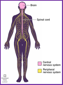
Peripheral nervous system branches out from central nervous system to reach every part of the body. Cleveland Clinic
Sensory receptors help us make sense of the world. In response to stimuli — both from within the body (interoception) and from the surrounding environment (exteroception) — sensory receptors convert stimuli into nerve impulses (neurons) and transmit them up the chain. The brain receives, sorts, interprets, and returns transmission via nerve fibers back to the source for an appropriate response — or no response.
Sensory receptors are located in our sense organs — ears, eyes, mouth, nose, skin — in the tissues and organs of our peripheral nervous system (PNS), and in the brain and spinal cord of our central nervous system (CNS). Different types of stimuli are sensed by different types of receptors — including in body systems responsible for senses we rarely consider: balance and movement, temperature and pain.
Sensory Transduction at the Bakery
I enter a bakery. My auditory sense registers the tinkling bell on the door, while my ocular and olfactory senses go to war for my attention. I see beautiful breads, cookies, and fruit tarts. But the smells emanating from the ovens in the back are even more alluring. My vestibular sense allows me to nod to the counter person’s welcome, as my visceral sense tells me my stomach needs immediate sustenance.
My body’s sensory system has gone haywire on my visit to the bakery. A collision of differentiated sensory receptors are simultaneously detecting and receiving these stimuli. External: bakery sounds, sights, smells. Internal: hunger, desire, anticipation. Through a process called sensory transduction, the stimuli are “transduced” or converted into electrochemical signals and conveyed by nerve impulses, called neurons, from the PNS to the CNS for response.

Receptor and Generator Potentials
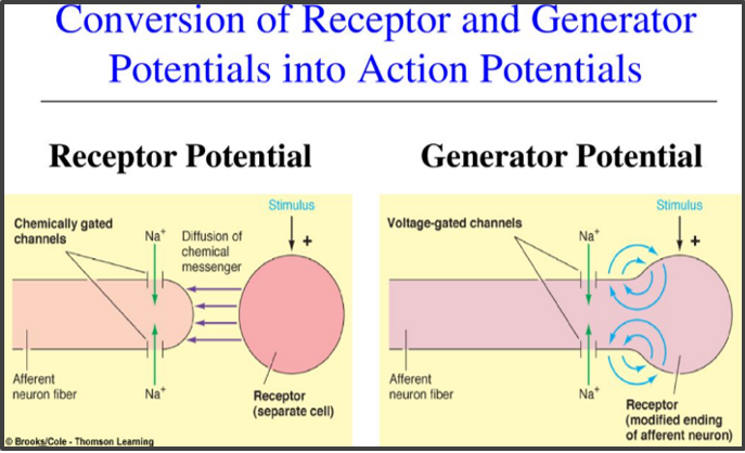
Receptor Potentials and Generator Potentials: Sensory information begins when a stimulus is converted into an electrical signal. This diagram shows two ways that process can occur: through a separate receptor cell that communicates with a sensory neuron (receptor potential) or directly within the sensory neuron’s own ending (generator potential). In both cases, the signal can trigger an action potential that carries information to the brain. From: Juliet Oliver, “The Peripheral Nervous System: Afferent Division.” Graphic: Brooks/Cole—Thomson Learning
First, a warning about the word potential. Researchers toss it around like it means cat or car. To understand it, you can’t think of the meaning in your head. It’s not about one’s ability or promise.
Science uses the term to replace the lack of reliable measures for something that may or may not happen. Potential is an accepted term for the temporary change in electrical charge across a cell’s membrane.
Potentials are often used to describe electrical or chemical signals in the body: receptor (or generator) potential, graded potential, and action potential.
When a sensory receptor is activated (by light, touch, sound), it creates a small, local change. This is called a receptor (or generator) potential — a type of graded potential, meaning its strength can vary. If that change becomes strong enough (like it’s given the grade of A+), it crosses a threshold and triggers a larger, all-or-nothing signal: an action potential, which carries information along a nerve cell.
So in this context, a potential is not a prediction — it’s a measurable shift that may or may not lead to something bigger.
Clear? Probably not if, like me, this is a first encounter with potentials — but it’s less important to understand potentials than to see how biology is working at the cellular level in transmitting sensory information. Here’s another take:
Neuron-to-Neuron Transmission
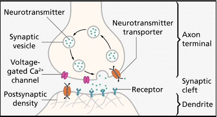
Structure of a Chemical Synapse: Key structures involved in communication between neurons. Wikipedia
Quick review from prior post: Action potentials (electrical signals) pulsate along the neuron’s axon to the presynaptic (before the synapse) or transmitting axon terminal. There, pouches of synaptic vesicles convert (transduce) electrical-to-chemical signals (neurotransmitters), which are released into the synaptic cleft. They bind onto the postsynaptic (after the synapse) or receiving dentrites of the next neuron in the chain. There, the chemical signals are reconverted into action potentials to once again travel the axon. All the way to the brain.
The reverse process is similar, but from the brain it’s motor neurons that do the conveying to the destination (sensory organs, glands, organs).
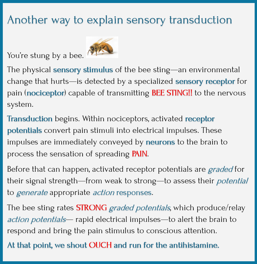
What’s remarkable in this process is the “electrical-chemical-electrical” transmission of impulses neuron-to-neuron, from the PNS to the CNS. The bee sting traveling in the spinal cord as electrical messages leaps across synapses as chemical signals and back into electrical signals to propagate the message further within each successive neuron.
- Due to the stinging pain, the chemical substance released into the synapse is probably Substance P (a neuropeptide that transmits pain signals), along with endorphins (not directly released by the pain stimulus, but as the body’s natural pain relievers).
- Through transduction and transmission, the pain message has reached the brain. The brain fires more action potentials when it’s excited. Action potentials are all or none: a neuron fires an action potential or it won’t.
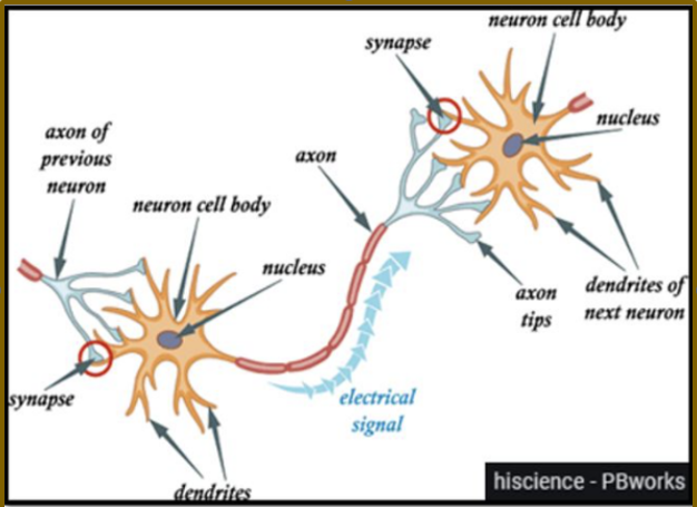
Structure of a Neuron: Major structures of a neuron and the direction of signal transmission. Psychopedia


- While sensory neurons traveled to the brain, motor neurons travel back to the PNS, to the site of the bee sting, sending electrical signals along nerve fibers and communicating between neurons as they did on the way to the brain. You are cognitively, consciously alert to your pain and deserving of sympathy.
The bee sting example shows how the nervous system responds — the whole thing taking milliseconds. But with pain, there’s also a shortcut: from stimulus to spinal cord and back, bypassing the brain, for faster response time. An example of this is stepping on a sharp rock and withdrawing the foot before the rock penetrates the skin.
Sensory receptors transduce stimuli that activate receptor potentials, transmitted as low-or-high graded potentials to the CNS/brain to generate action potentials. —Wikipedia
- Weakly graded potentials don’t generate action potentials — making it unlikely sensory stimuli reach our consciousness.
- Strongly graded potentials (like in the bakery) produce and relay (generate) action potentials, which travel from the PNS to the CNS.
- Once in the CNS, action potentials are integrated with other sensory information — and sometimes higher cognitive functions — to become a conscious perception of the stimuli.
- Central integration can lead to a motor response. It does. I buy and eat the pastries. My happiness and satiety trigger further sensory stimuli, and the process continues.
Graded Action Potentials Have Real-Life Consequences
Most graded potentials never reach conscious perception. If I was on my phone walking past the bakery, I might have missed taking in the alluring smells of freshly baked bread. Sensory stimuli entered my olfactory sensory receptors, but did not penetrate my consciousness — so I was denied my morning treat.
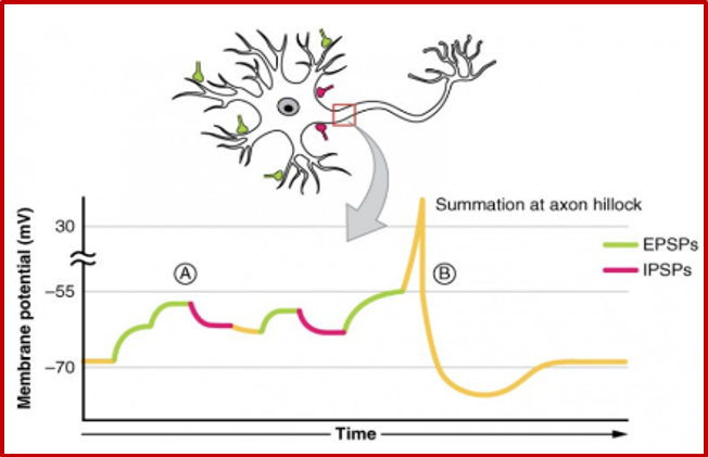
Postsynaptic Potential Summation: A neuron constantly receives messages that either encourage it to fire or hold it back. These incoming signals are added together, or summated, to produce a net effect. At A, excitatory signals combine and push the neuron closer to firing. At B, excitatory and inhibitory signals interact, and the balance between them determines the neuron’s response. Lumen Learning
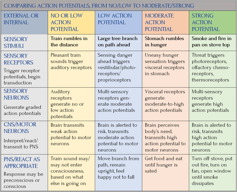
In this chart, I created scenarios to illustrate gradations in the sensory process:
Sensory Organs Avert a Crisis
- Sight: Eyes send signals that smoke is building on the stove
- Smell: Brain perceives something nearby is burning
- Taste: Taste chemoreceptors on taste buds bind chemical compounds, allowing you to taste
- Hearing: Hair cells in the inner ear convert sound waves into electrical signals
- Touch: Nerves in your hand sending a message to the brain registers the stove as “hot”
Sensory receptors fire off thousands of signals at a time, take milliseconds to do their work, and flood our brains to determine and prioritize responses, if any. The brain determines whether to react to transmitted signals.
Rewards and Our Pursuit of Them
The sensory system is hardwired into the body’s life force. But so, too, is something we may not think as much about: how hardwired we are to pursue, attain, consume, and evaluate rewards that motivate us in life. Rewards are the appealing, motivational parts of a stimulus (any object, event, activity, situation, or person). Being singled out for a job well done, smelling food being prepared in the kitchen, or a positive social experience like laughter can all activate the reward system, promoting feelings of wellbeing and social connection. At whatever level, from small acts of kindness to receiving the Nobel Peace Prize, we desire these rewards. Rewards can elicit positive reinforcers and vice versa — positive reinforcers are rewarding.
Scientists in almost every discipline study reward systems in the animal kingdom. They tell us about our evolutionary choices and primitive drives to survive and propagate. Animals are motivated to repeat pleasure-seeking behavior: to sate hunger/thirst, to hunt prey or, if prey themselves, go into camouflage mode, run, climb trees, dig holes. And to attract mates.
The Reward and motivation System
One of the brain’s most important dopamine networks is the mesocorticolimbic system, often called the brain’s reward system. It originates in the ventral tegmental area (VTA) and connects with regions involved in motivation, learning, decision-making, and emotional regulation, including the nucleus accumbens and the prefrontal cortex (PFC). Rather than simply producing pleasure, this network helps the brain identify what is important, anticipate rewards, learn from experience, and direct attention and behavior toward meaningful goals.
Sources for this section: Neuroscientifically Challenged, Research Gate, Simply Psychology, PubMedCentral/NIH, and Psychology Today
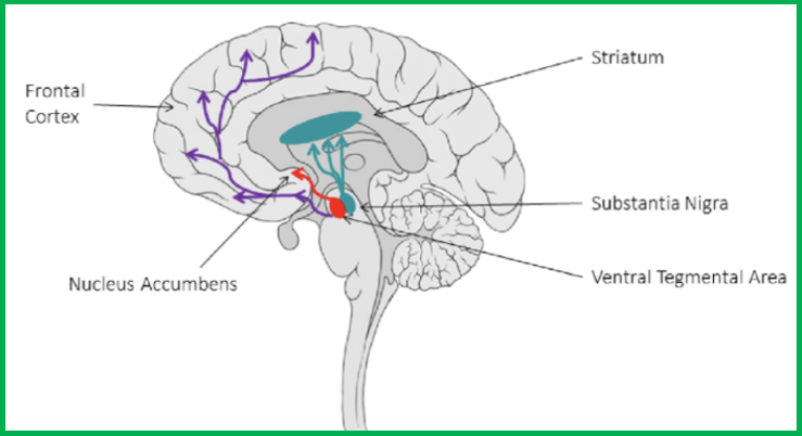
Major Dopamine Pathways in the Brain: Dopamine travels through several pathways that support different functions. In this diagram, the purple pathway is involved in thinking, planning, and self-regulation; red contributes to reward, motivation, and learning; and blue-green helps coordinate movement. Together, these pathways influence how we think, feel, learn, and act. Image: Wikipedia
Reward Cognition, Anticipation, and Loops
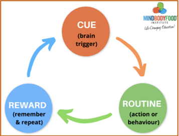
Source: Mind Body Food Institute
Reward cognition triggers the reward system to seek out potentially pleasurable, motivating experiences. Science recognized dopamine as essential to feelings of pleasure back in the 1950s, but more recently has found it’s also essential to reward anticipation — pricking up our senses to detect cues from the environment or internal sensations and then sharpening our focus on what we have to do, before taking action. In reward anticipation, our entire body becomes goal-directed. Motor preparation allows us to move toward acquiring the reward. We fire up our cognitive and emotional engines to ready ourselves for some kind of beneficial experience, either to participate (passive) or to perform an act (active). When it’s over, we can assess whether the received reward matched the anticipated one.
Anticipating rewards isn’t only linked to dopamine release on the dopamine pathway, but also involves the anterior cingulate cortex (ACC), which processes both limbic system (emotion) and PFC information. Reward seeking is a mind-brain-body affair.
World-renowned American psychologist/ inventor B.F. Skinner (1904-1990) pioneered the study of operant conditioning and behaviorism (similar to the classical conditioning of Ivan Pavlov’s dogs). Skinner developed what he called the reward loop (also called compulsion or habit loop), a three-part, self-perpetuating circuit. Cue triggers behavior, routine is the action we perform, and finally reward is the positive reinforcement that motivates us to repeat the action. Due to synaptic plasticity in the circuit (ability to modify strength of neural connection based on new experience), reward loops are the basis of habit formation, both positive and negative. Repeated positive reinforcement strengthens cue-to-routine connections, making behavior seem automatic (constantly checking social media). But in drug addiction, some substances can over-activate the reward loop, leading to compulsive-seeking behavior.
B.F. Skinner, Very Well Mind
More Pos/Neg Facts About our Reward Systems
Our brain’s reward system is exceedingly complex and its affects are so bound up in who we are and how we behave that we couldn’t possible know what life would be like stripped of this particular innate gift. I close with more things to know about our reward system.
According to researcher Kent Berridge, there are two systems, the “wanting” and the “liking,” and these two systems are complementary. Dopamine is part of the wanting system. It propels you to take action. The liking system makes you feel satisfied and therefore pause your seeking. But the dopamine wanting system is stronger than the liking system. You tend to seek more than you are satisfied. You can get into a dopamine loop. If your seeking isn’t turned off at least for a little while, then you start to run in an endless loop. —Susan Weinschenk, PhD, “The Dopamine Seeking –Reward Loop”
- Reward learning, a subtype of associative learning, is the association of positive stimuli, actions, and contexts (the reward) with positive outcomes. When we modify our behavior based on the outcomes, reward cognition strengthens the neural pathways associated with that behavior. In this way, we’re encouraged to repeat the desired behavior
- In associative learning, the neural connections between two or more stimuli — creating a mental association in memory — are both positive and negative. Reward learning is mostly positive, unless it leads to negative outcomes, like bad habits.
- Reward learning is based on operant conditioning — we’re wired to seek pleasure and avoid pain. Skinner’s theorized that we learn through reward/punishment, modifying behavior through consequences. Positive punishment decreases a behavior by adding an aversive stimulus; negative punishment decreases a behavior by removing a positive one. A parent praising a child for cleaning their room is positive reinforcement. Ignoring a child who won’t clean their room is negative punishment — removing approval or praise. Both responses encourage positive behavior.
- Positively valenced / pleasurable emotions that determine the emotional effect of an event, object, or situation are on a positive valence continuum — from intrinsic good, happiness, joy, attractiveness, or appeal to ecstasy and euphoria.
Coming Up Next

Over the pages of this blog, I hope this rudimentary understanding of the neurobiology of how our nervous system works will be a solid foundation to understand what’s atypical about the neurodivergent brain. So far, I’ve covered sensory transmission, action potentials, transduction, and transmission across synapses, And our body’s reward system, which motivates us consciously/unconsciously, positively/negatively, in small/large ways. In Post 5: Sensory Receptors are the Body’s Cellular Plan, I’ll continue with sensory transmission by describing sensory receptors and how the sensory system communicates within the brain and out to the eyes and ears, the limbs and body in balance, in space.
Copyright ©2026 Jan Swan