On Neurodivergence and Otherness
Show Table of Contents
INTRO
1. On Neurodivergence and Otherness: An Introduction
SENSES AND SENSORY SENSITIVITIES
2. Senses Count
3. Neurobiology for Dummies
4. Sensory Transmission and our Reward System
5. Sensory Receptors are the Body’s Cellular Plan
6. A Synthesis: Sensory Systems and our Emotions — Part I
7. A Synthesis: Sensory Systems and our Emotions — Part II
8. Sensory Disorders and Sensitivities
9. Etan’s Story
10. Synesthesia: Difference, But Not Disorder
11. Synesthesia, Creativity, Artistry — Part I
12. Synesthesia, Creativity, Artistry — Part II
AUTISM AND THE NEURODIVERSITY MOVEMENT
13. From “Mental Defectives” to Autism Spectrum Disorder
14. Changing Conception of Autism
15. Autism Diagnoses and Behavior Patterns
16. Autism Treatments that Help
17. Early Start Autism Treatment: A Case Study
18. Neurodivergence and the Neurodiversity Movement
19. Neurodiversity Takes Flight
ADHD
20. ADHD and Neurodevelopmental Disorders
21. ADHD: A Preponderance of Risk Factors and Symptoms
22. ADHD: Inattentive, Impulsive…and Hyperactive?
23. ADHD: Named, Renamed, Still Needs a New Name
24. ADHD: Treatment and Coping Skills for All Ages
LGBTQ+
25. Neurodiversity and the LGBTQ+ Fight
26. LGBTQ+ Identity and Expression
27. LGBTQ+ and Mental and Behavioral Healthcare
ON LANGUAGE
28. Language Matters In and Around Neurodiversity
29. Neurodivergent Language Difficulties
30. Disability-Inclusive Language Guidelines
ON CREATIVITY AND GIFTEDNESS
31. Neurodiversity and Creativity
32. Giftedness is a Piece of Neurodivergence
SELF-IDENTITY
33. Self-Identity: The Cornerstone of Neurodiversity
34. Early Theories of Self-Identity Formation
35. Contemporary Theories of Self-Identity Formation
36. Authenticity and the Search for Self
37. Self-Schemas and Neurodivergence
38. Self-Labeling and Parts Work
39. Complexity, Clarity, and Self
IMPROVING LIFE FOR NEURODIVERGENT PEOPLE
40. Empathy Recognizes and Navigates Difference
41. Reducing Neurotypical-on-Neuroatypical Conflict – Part I
42. Reducing Neurotypical-on-Neuroatypical Conflict – Part II
43. Communicating Across the Neurospectrum – Part I
44. Communicating Across the Neurospectrum – Part II
45. Neurodiversity: Advocacy and Education
46. Neuroinclusion in the Workplace
47. A Neurodiverse Lifestyle
IN CONCLUSION
48. In Conclusion: Neurodivergence and Inspiration
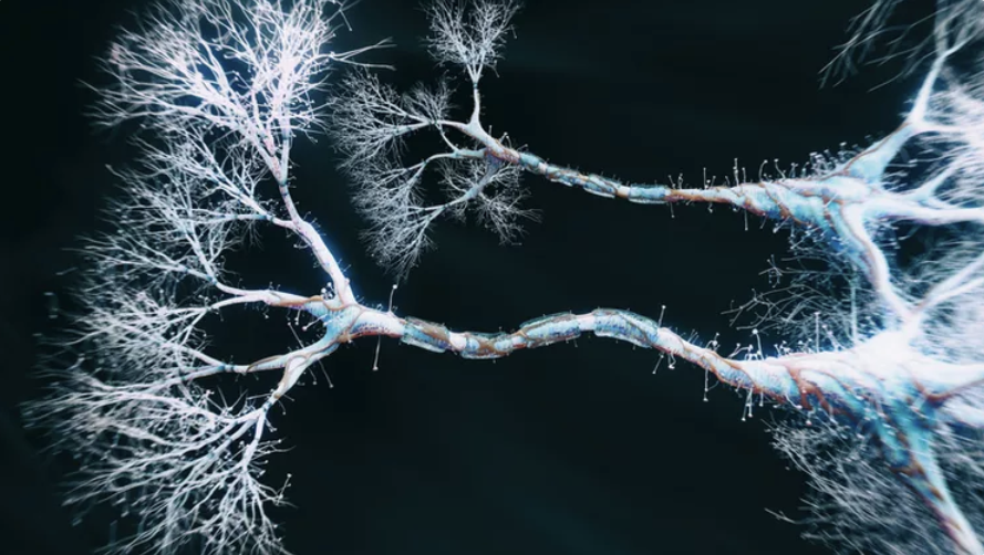
Picture of axons, Science Direct
Do I have to give the “…for Dummies” books credit for the title of this post? For Dummies is an incredibly successful international franchise of “non-intimidating guides for readers new to topics covered.” And with a nod to a great idea, I’m planning to make science — to the extent I can — more digestible throughout this and other series.
I do this out of conviction. We can’t understand mental health/neurodivergent challenges without some insight into the neurobiophysiological sources of difference.
The mind-body-brain connection is key, but blurry lines separate these distinctions. In these next posts, I’ll explain how nerve cells (neurons) interact and communicate (via synapses) within the body’s nervous system — toward processing information and influencing how we think and act.
A foundation in the sensory system’s underlying biology helps explain why most of us are neurotypical (NT) and some of us are neurodivergent (ND). In places you’ll need to draw your own conclusions, because science is making progress but doesn’t yet have all the answers.
I’ve shored up support for why we need to know some science by consulting Harvard University’s Mind/Brain/Behavior Interfaculty Initiative (MBB), a group of interdisciplinary researchers who aim to “elucidate the structure, function, evolution, development, and pathology of the nervous system in relation to behavior and mental life.” Neuroscientist Jeff Lichtman, et al., explains:
Photo: Baby Memorial Hospitals
Neurobiology is concerned with uncovering the biological mechanisms by which nervous systems mediate behavior. Over the past half century, much of neurobiology has focused on…the structure and physiology of nerve cells (neurons)…and the functional contacts (synapses) made between neurons. How individual nerve and receptor cells generate, carry, and transmit electrical and chemical signals is now well understood….
As we move into the 21st century, increasing attention is being given to integrative or systems neurobiology—the study of aggregates of neurons and functional circuits. How do assemblies of neurons give rise to the behaviors we associate with higher brain functions, from perception and control of movement to learning and memory? —Jeff Lichtman, et al., Mind/Brain/Behavior Interfaculty Initiative, Harvard University
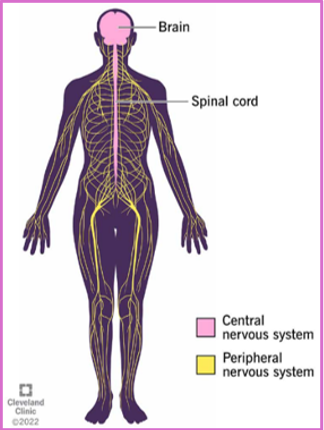
Our Nervous System
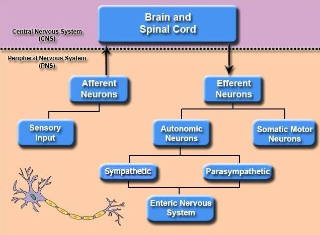
Organization of the Nervous System. Image generated at BYU-Idaho
The sensory system is a specific part of the global nervous system. Whether presiding over voluntary or involuntary actions, the entire nervous system works on the transmission of neurons from the body’s periphery to the brain and back to the periphery.
I discussed proprioception in the last post. Here’s an example of how we need to understand both the global and the specific, through the case of Christina:
Imagine you suddenly lost the ability to stand unless you looked down at your feet. What if your arms seemed to wander unless you kept an eye on them? This is the exact situation Christina found herself in one morning. She exclaimed: “Something awful’s happened, I can’t feel my body. I feel weird — disembodied.” Essentially overnight, Christina had lost all proprioception: the ability to sense the relative position of body parts. This integral part of her nervous system had suddenly deteriorated.Because the sense of body positioning is determined by three things — vision, balance organs, and proprioception, which she had lost — Christina learned to control her body with her eyes. But she found that she could do nothing without using her eyes. Her body would collapse into a heap the minute she closed her eyes.
Gradually over time, Christina learned to walk again and to function with the usual business of life, but only with great care…. She found there was no in between or gradual change with movement …. She states: “I feel my body is blind and deaf to itself … it has no sense of itself.” Christina’s case [shows] how the nervous system interprets the environment around us. —“Anatomy & Physiology,” Brigham Young University, Idaho

Photo: Outreach-rehab
According to multiple sources, the nervous system is made up of 7 trillion nerves — end to end that translates to 45 miles of nerves — with 86 billion neurons (revised down from 100 billion) and a 10-fold increase of glial (supporting) cells. An individual nerve contains thousands-to-millions of neurons, the large variation depending on size, function, and location in the body. However, a more interesting fact is that each neuron can make on average 7,000 synaptic connections to other neurons, meaning the adult brain ranges from 100 to 500 trillion synapses. The vast majority are in the brain and mostly in the brain’s cerebrum, but they are also located throughout the nervous system.
- Central nervous system (CNS) is the brain and spinal cord, consisting of neurons associated with central processing.
- Peripheral nervous system (PNS) connects the CNS to the rest of the body—sensory organs, visceral organs, glands, muscles—and consists of neurons associated with sensory input (afferent) and motor output (efferent).
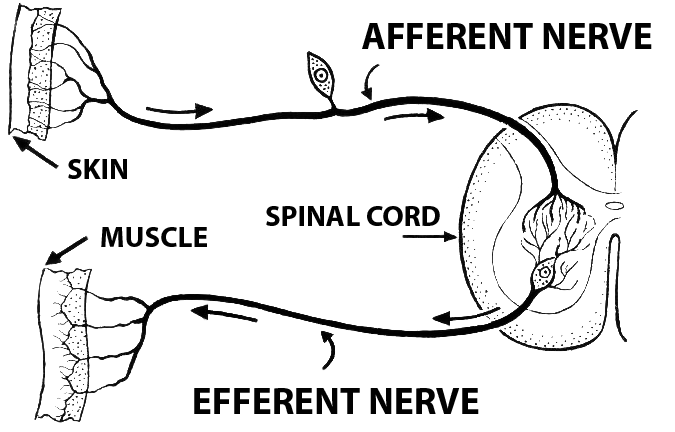
Afferent vs Efferent Neurons
Afferent or sensory neurons respond to all sensory stimuli and help us perceive the world around us. Afferent neurons are part of afferent nerve fibers carrying sensory information from points all over the body (PNS) to the spinal cord and brain (CNS). Afferent nerves are primarily found in sensory organs (eyes, ears, nose, tongue, skin) — essentially where sensory information is received from the external environment. But they’re also found in visceral organs (heart, stomach, lungs, etc.), where sensory neurons monitor the internal environment and organ systems and respond to pain, blood pressure, hunger, and inflammation.
Source: Wikipedia Commons
Just the opposite are efferent or motor neurons — part of efferent nerve fibers — which move messages away from the CNS and toward the PNS, specifically to stimulate motion in the muscles and glands. Efferent nerves can be further divided into the somatic nervous system (controls voluntary movement of skeletal muscle) and the autonomic nervous system/ANS (regulates involuntary functions).
Mixed nerves contain both sensory and motor fibers, carrying sensory information to the CNS and motor commands back to the PNS.
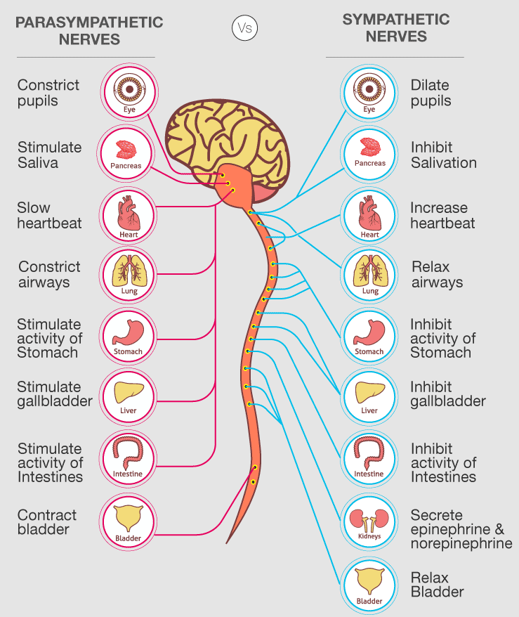
Autonomic Nervous System (ANS)
Part of the PNS, the autonomic nervous system (ANS) is made up of nerves that connect the spinal cord to the heart, stomach, intestines, and other visceral organs. The ANS controls and regulates involuntary physiologic processes, including heartrate, blood pressure, respiration, digestion, and sexual arousal. The ANS has three divisions:
- Sympathetic nervous system activates body processes that help in times of stress or danger — responsible for the body’s fight-or-flight response/FFR [covered in post 5, in the anxiety series].
- Parasympathetic nervous system is responsible for rest-and-digest body processes, including during FFR.
- Enteric nervous system controls the gastrointestinal tract. It runs independently or is modulated by the ANS. It contains more neurons than the spinal cord.
Source: BYJU’s Biology
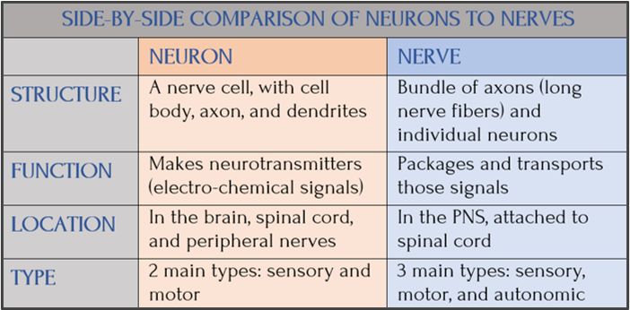
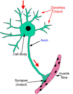
Neurons, Nerves, and Synapses
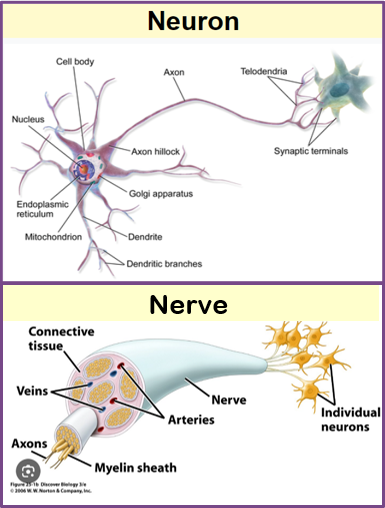
Neurons are “excitable nerve cells that fire electric signals called action potentials across a neural network in the nervous system” [Wikipedia]. Neurons send messages to other neurons via a specialized connection: the synapse. The long nerve fibers connecting neurons are called axons.
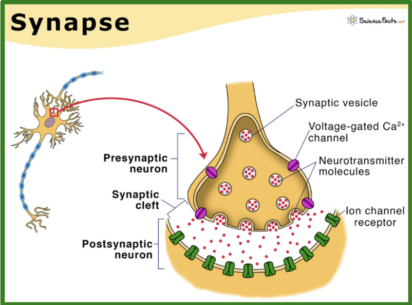
Neurons are sending and receiving messages all the time, much like the telephone game when you whisper from person to person to see how the message changes. Except in the nervous system, the message stays faithful to the original. Transmission of information via electrochemical impulses — neurotransmitters — crosses the synapse (synaptic cleft). So neurons before the synapse are presynaptic and after the synapse postsynaptic.
Synapses are created by two terminals (transmitting and receiving) at either end of the axon and the gap between (synaptic cleft). The presynaptic terminal converts electrical signals to neurotransmitters (chemical messengers), which are released into the synaptic cleft and picked up on the other side by receptor molecules on the postsynaptic terminal membrane (50 nanometers away).
This is hard to follow without a diagram. Think about ears, cell phone networks, and signals, and it starts to make sense. Here’s an example of message transmission between, say, neuron A and B:
- Neuron A: Presynaptic (transmitting) neuron releases electrochemical message (via neurotransmitter) from its “transmitting synaptic terminal” into the synaptic cleft and toward neuron B.
- Neuron B: Postsynaptic (receiving) neuron has receivers that latch onto neuro-transmitted impulses and send them along the axon to the neuron’s transmitting end — which repeats to send message to neuron C, until reaching the journey’s destination (the brain).
Synaptic transmission. Source: Science Facts
In sum: the message begins in neuron A as an electrical impulse, gets converted before the synapse to chemical signals (neurotransmitters) and then, reaching neuron B at the receiving end, is reconverted into electrical signals and carried along the axon to neuron C — until reaching the brain.
This is hard to follow without a diagram. Think about ears, cellphone networks, and signals, and it starts to make sense. An example of message transmission between, say, neuron A and B:
Axon terminal, creating a synapse. Source: Science Direct
Brain Networks
At the risk of over-simplifying how the brain works, we need to understand it to see just how many brain networks are responsible for how we function, process complex amounts of information, manage our emotions, and control our behaviors to be appropriate for the tasks ahead.
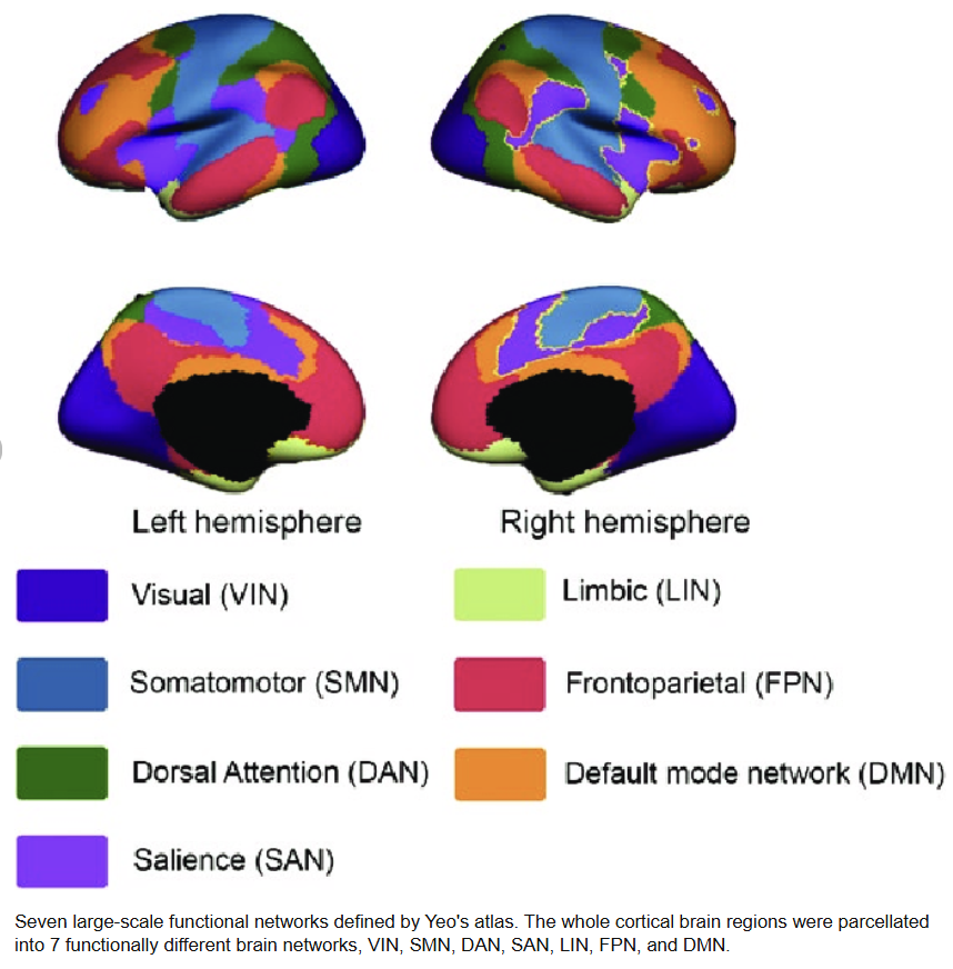
Source: ResearchGate
Our interconnected brain networks spread across different brain regions and work together to perform these complex mental processes. They’re not static but dynamically switching to meet the need. The strength and pattern of neural connections between brain regions within a network are crucial for its functioning — as well as between the brain networks. In this way, the power of the full brain is brought to each specialized area, making us effective and efficient and capable of parallel processing.
Different networks are more closely involved with cognitive control/executive function (EF) processes, like memory, language, decision-making, visual perception, and spatial navigation, while others are responsible for processing emotions, linking emotional states to cognitive processes.
Sources: Above information was drawn from Science Direct, PubMed/NIH, Understood, The Lancet, and Research Gate
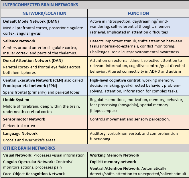
Brain Networks and Impairment
Psychiatric research increasingly points to interacting brain networks whose altered functioning can contribute to both mental health and neurological conditions. Depression, anxiety, and PTSD, as well as typically lifelong, brain-based conditions, such as sensory processing differences, autism, and ADHD, which exist along a range of impairment.
One way to understand this is to think about how well the brain’s networks function within themselves and in coordination with one another. When these systems are not working smoothly, mental processes can become slowed, blocked, or misdirected — making it harder to think clearly, regulate emotion, stay organized, or relate to others.
PTSD, for example, has been linked to disrupted coordination between the brain’s core networks for internal focus (the default mode network/DMN), goal-directed attention (the central executive network/CEN), and salience detection and switching. Similarly, research in ADHD and autism points to differences in connectivity within and between networks involved in attention and salience — including the default mode, dorsal attention, and salience networks — though the specific patterns of disruption are not the same.
These brain-based conditions are not about laziness, being weird, trendy, or less intelligent, but they can create real challenges that may require support and treatment. There are effective approaches that help people function, adapt, and do well in their lives
Coming Up Next
In Post 4: Sensory Transmission and our Reward System, I’ll cover a crucial step in sensory transmission, called transduction — the conversion of sensory stimuli to electrochemical impulses passed through neurons on their way to the brain. I’ll describe how, through this conversion process, impulses are “graded” weak-to-strong to guide the brain in finding an appropriate response. I’ll introduce a word much encountered in sense science: “potentials” or stand-ins for the potential of an action and the lack of measurable specifics. The next post will conclude with a cursory look at a fascinating but often overlooked body phenomenon: our reward system and the why and how of motivation.
Copyright ©2026 Jan Swan